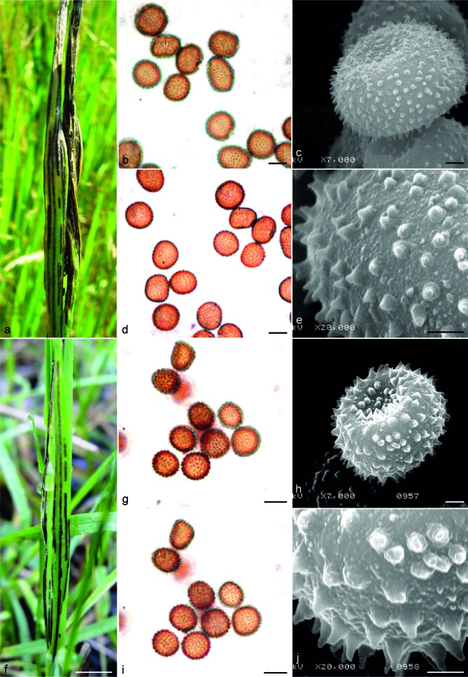
Fig. 5.
Sori and spores of Ustilago bromina (a–e) and U. striiformis s.str. (f–j). a. Sori of U. bromina (HAI 4600); b, d. teliospores seen by LM, superficial, and median views respectively (S F163740); c. teliospore seen by SEM (S F163740); e. surface of teliospore seen by SEM (S F163740); f. sori of U. striiformis s.str. (HAI 4608); g, i. teliospores seen by LM, superficial, and median views, respectively (S F163771); h. teliospore seen by SEM (S F163771); j. surface of teliospore seen by SEM (S F163771). — Scale bars: a, f = 1 cm; b, d, g, I = 10 μm; c, h = 2 μm; e, j = 1 μm.

