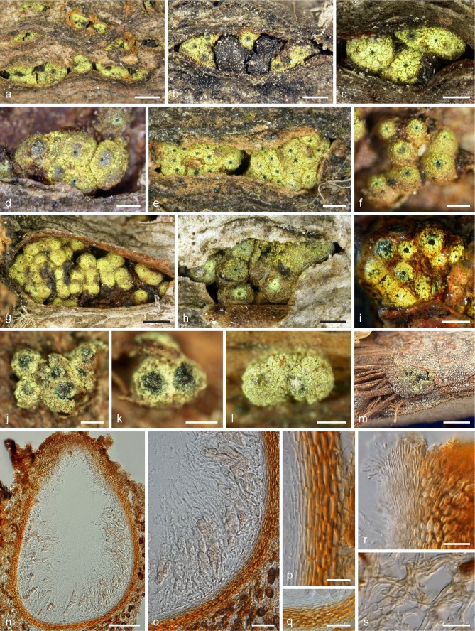
Fig. 3.
Thyronectria rhodochlora, stromata and ascomata. a–h, j–m. Dry stromata in surface view (a. habit; b. with pycnidia of a Diplodia sp.); i. stromata in 3 % KOH after rehydration; n. perithecium in vertical section; o. lower lateral portion of a perithecium showing asci and apical paraphyses in section; p–r. peridium in section (q. basal region; r. ostiolar region with periphyses and scurf); s. stroma hyphae (n–p, r: in 50 % glycerol; q, s: in lactic acid). a, e, i, n–s: epitype WU 31656 (NP3); b, c, g: WU 32152 (NP7); d, f, h: WU 31654 (NP1); j, k: lectotype (PC); l: isolectotype (K); m: holotype of T. patavina (PAD). — Scale bars: a, m = 1 mm; b, e, h, i = 0.4 mm; c, d, f, j–l = 0.3 mm; g = 0.7 mm; n = 100 μm; o, q = 30 μm; p, r, s = 15 μm.

