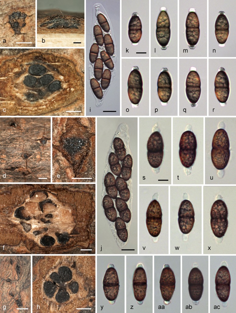
Fig. 12.
Phaeodiaporthe appendiculata. a, d, e, g. Ectostromatic discs and ostioles in surface view; b. pseudostroma in vertical section; c, f, h. pseudostromata in transverse section, showing perithecia, whitish to brownish entostromata and faint blackish marginal zones; i, j. mature dead asci with apical ascal ring in i; k–r. living ascospores with blunt gelatinous appendages; s–ac. dead ascospores with blunt gelatinous appendages with i, k–r, y–aa in water and j, s–x, ab, ac in 3 % KOH (a–c, i. WU 32449 (epitype); d, e, f, j, s–x. B 700021801 (holotype of Diaporthe appendiculata); g, h, y–ac (lectotype of Phaeodiaporthe keissleri); k–r. WU 32448). –– Scale bars: a–c, e, f, h = 0.5 mm; d, g = 1 mm; i, j = 20 μm; k–ac = 10 μm.

