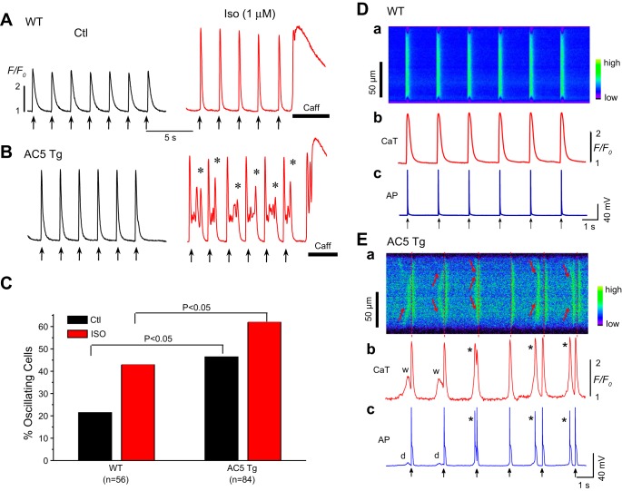
Fig. 7.
Spontaneous Ca2+ waves and triggered activities (TAs) in AC5Tg myocytes. A: representative Ca2+ fluorescence recordings in the absence (Ctl) and presence of isoproterenol (1 μM) in a WT myocyte. B: representative Ca2+ fluorescence recordings in the absence (Ctl) and presence of isoproterenol (1 μM) in an AC5Tg myocyte. Prominent Ca2+ aftertransients (asterisks) were observed in AC5Tg myocytes after isoproterenol challenge. C: incidence rate of Ca2+ oscillations and waves in WT and AC5Tg myocytes in the presence and absence of 1 μM isoproterenol. Myocytes used in observations were from 3–5 mice in each group. E: simultaneous line-scan image along the long axis of the myocyte (Ea), Ca2+i transients (Eb), and APs (Ec) from an AC5Tg myocyte in the presence of isoproterenol (1 μM). Aberrant spontaneous Ca2+ waves (indicated by the arrows in Ea, and w's in Eb) causing delayed afterdepolarizations (DADs) (d) and triggered beats (*) in Eb and Ec. Pacing marks are indicated by the arrows. D: same as E, except from a WT myocyte. Neither apparent Ca2+ waves nor triggered activities were observed in this specific cell.

