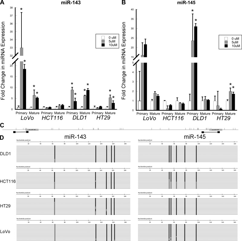
Fig. 2.
Primary and mature miR-143 and miR-145 expression and DNA methylation in colon cancer cells. A and B: real-time PCR measurement of expression of mature and primary miR-143 and miR-145 following 5 consecutive days of treatment with 0, 5, or 10 μM 5-aza-2′-deoxycytidine (5-AZA). C: primer positions (black boxes) relative to miRNAs (gray boxes) and CpG locations (vertical bars) for sequences of bisulfite-treated DNA around genes encoding miR-143 and miR-145. D: sequencing for the indicated loci in DLD-1, HCT-116, HT-29, and LoVo colon cancer cells after bisulfite conversion and PCR amplification. Each line represents a single clone. Black boxes, methylated CpG sites; gray boxes, unmethylated CpG sites.

