
Figure 4.
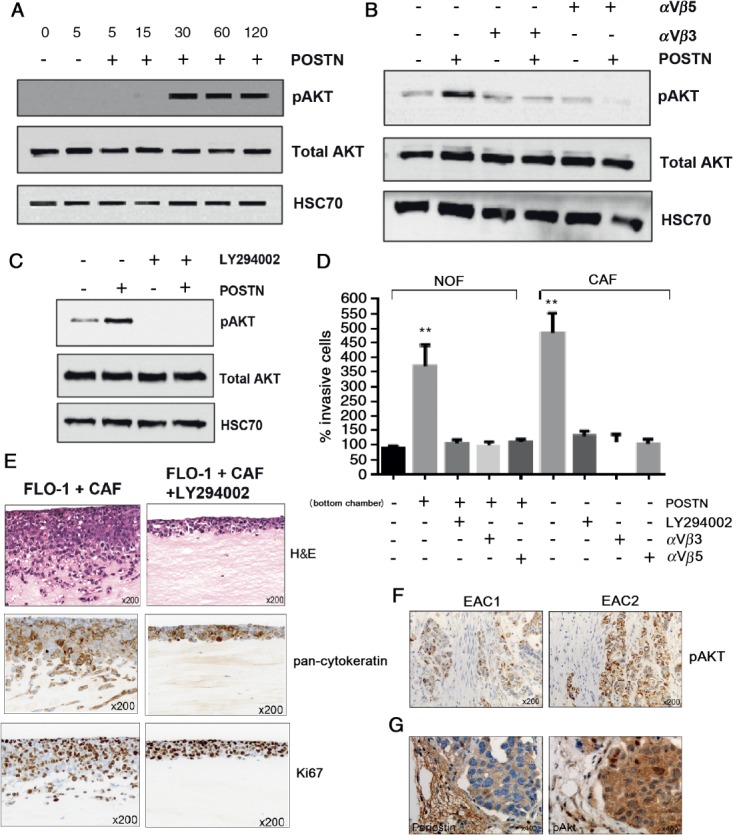
Periostin-dependent invasion of EAC cells is suppressed through inhibition of αvβ3 and αvβ5 integrins and PI3K. (A) Western blot for total and phospho-Akt in FLO-1 up to 120 min after stimulation with recombinant periostin. (B) Western blot for total and phospho-Akt in FLO-1, 30 min after periostin stimulation, in the presence or absence of blocking antibodies against integrins αvβ3 or αvβ5, as indicated. (C) Western blot for total and phospho-Akt in FLO-1, 30 min after periostin stimulation, in the presence or absence of the PI3K inhibitor LY294002. (D) Transwell invasion assays of FLO-1 towards NOFs or CAFs conditioned medium under varying conditions. Invasion towards NOF-conditioned medium is used as the internal control. Periostin was added to the NOF-conditioned medium and either LY294002 or blocking antibodies against integrins αvβ3 or αvβ5 were added to the FLO-1 cells, as indicated. (E) Organotypic models of FLO-1 + CAFs in the presence or absence of LY294002, including staining by H&E, pan-cytokeratin and Ki67. (F) Immunohistochemistry for p-Akt in representative human EAC tumours with strongest staining adjacent to CAFs. (G) Immunohistochemistry for periostin and p-Akt in tumour xenografts containing OE33 and CAFs