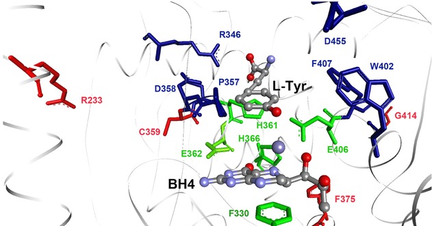
Figure 4.

A closer view of the active site of human TH. In green are the iron coordinating triade (His361, His366, and Glu406) and the BH4 binding Glu363 and Phe330. In blue are the substrate interacting residues Arg346, Asp358, Pro357, Trp402, Phe330, and Asp455 (based on the structure of the catalytic domain of human PAH with BH4 and 3-(2-thienyl)-l-alanine (PDB 1KW0)). Residues Arg233, Cys359, Phe375, and Gly414 are shown in red.
