
Figure 4.
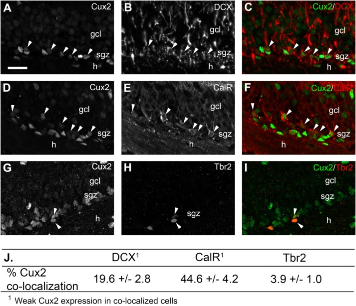
Cux2 is expressed in neuroblasts and immature granule neurons. A–C: Cux2 (A, C, green) staining in DCX+ neuroblasts (B, C, red) in the SGZ at P21. C: The Cux2/DCX co-stained cells (arrowheads) typically displayed lower levels of Cux2 protein relative to adjacent SGZ cells. D–F: Cux2 expression (D, F, green) in Calretinin+ immature neurons (E, F, red) arrowheads). F: Co-stained cells expressed lower levels of Cux2 (arrowheads). G–I: Limited Cux2 expression (G, I, green) in Tbr2+ transit amplifiers (H, I, red; arrowhead). J: Quantification of Cux2 co-localization with DCX, CalR, and Tbr2. Most Cux2+ (44.6%) cells in the SGZ were CalR+ immature neurons. Cux2 co-staining in DCX+ and CalR+ cells displayed low levels of Cux2 expression. Scale bar: A, 25 µm. Abbreviations as in Figure 3; CalR, Calretinin. [Color figure can be viewed in the online issue, which is available at http://wileyonlinelibrary.com.]