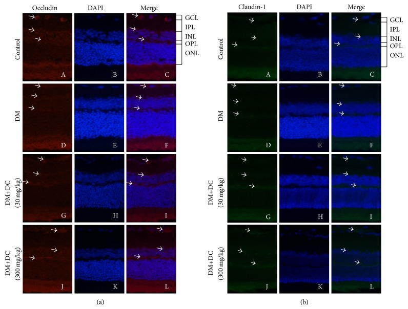
Figure 3.
The expression of occludin and claudin-1 in retinas. (a) The representative pictures of retinal immunofluorescence staining of occludin (A, D, G, and J) and DAPI (B, E, H, and K). Merge of occludin- and DAPI-stained images (C, F, I, and L). (b) The representative pictures of retinal immunofluorescence staining of claudin-1 (A, D, G, and J) and DAPI (B, E, H, and K). Merge of claudin-1- and DAPI-stained images (C, F, I, and L) (original magnification ×400).

