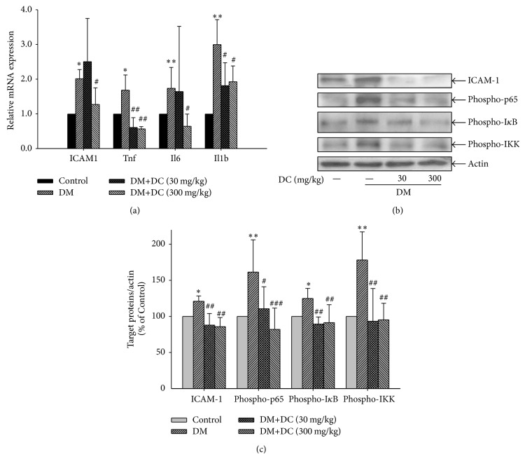
Figure 4.
Effects of DC on the expression of ICAM-1, TNFα, IL-6, IL-1β, and the activation of NF-κB signaling pathway. (a) Retinal mRNA expression of ICAM-1 (ICAM1), TNFα (Tnf), IL-6 (Il6), IL-1β (Il1b). Data are expressed as means ± SD (n = 10 for control, n = 7 for DM, n = 9 for DM+DC 30 mg/kg, and n = 8 for DM+DC 300 mg/kg). * P < 0.05, ** P < 0.01 compared to control; # P < 0.05, ## P < 0.01 compared to DM without DC. (b) Retinal expression of ICAM-1 and phosphorylated p65, IκB, and IKK. ICAM-1 and phosphorylated p65, IκB, and IKK are detected by immunoblotting using specific antibody. Results represent at least three repeated experiments. (c) Quantitative densitometric analysis of ICAM-1 and phosphorylated p65, IκB, and IKK. Data are expressed as means ± SD (n = 10 for control, n = 7 for DM, n = 9 for DM+DC 30 mg/kg, and n = 8 for DM+DC 300 mg/kg). * P < 0.05, ** P < 0.01 compared to control; # P < 0.05, ## P < 0.01, and ### P < 0.001 compared to DM without DC.

