
Figure 4.
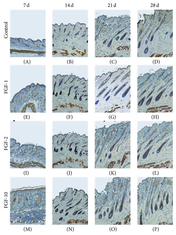
The expression of Shh after topical application of FGFs. Longitudinal sections of the dorsal skins from each group were stained for Shh by immunohistochemistry (brown staining). Digital photomicrographs were taken from representative areas at a fixed magnification of 100x.