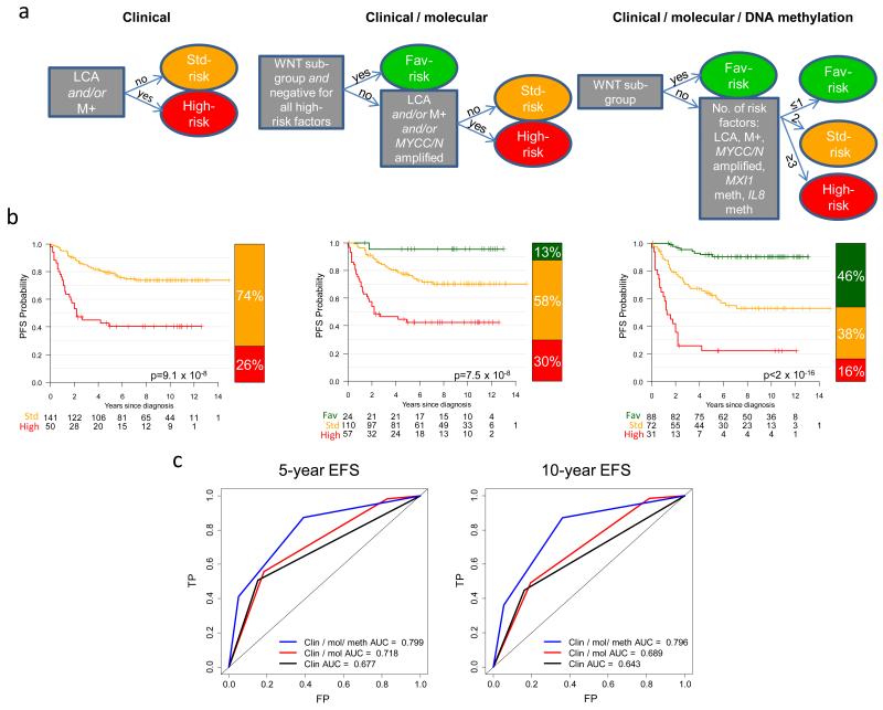
Figure 5. Risk-stratification using DNA methylation markers in 191 medulloblastoma patients aged 3.0 to 16.0 at diagnosis.
a. Current clinical (first panel) and clinical / molecular (second panel) models were assessed alongside a novel model incorporating DNA methylation biomarkers (third panel), and used to derive Kaplan-Meier plots (b). LCA, large-cell/anaplastic pathology; M+, metastatic disease; MXI1 and IL8 meth, methylated (β≥0.67). Low-risk, green; standard-risk, amber; high-risk, red. P values from log-rank tests are shown. c. Time-dependent ROC curves for all three models at 5 and 10 years. The area under the curve (AUC) is shown for each model. FP, false positive rate; TP, true positive rate. Fav, favorable.

