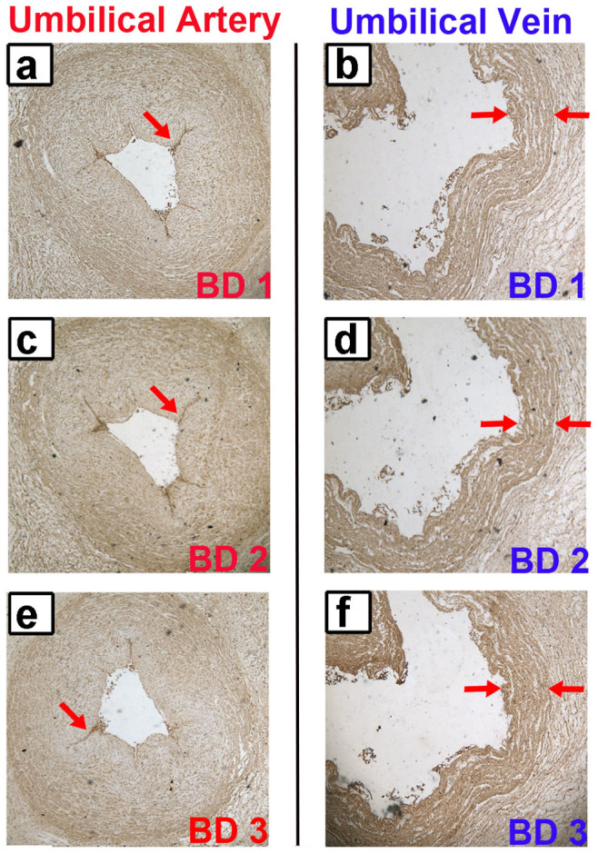
Figure 6. Staining of the BD autoantigens in human umbilical tissues.

(a, b) The staining of the BD1 patient in human umbilical artery and vein was analyzed by immunohistochemistry method. (c, d) The staining of the BD2 patient in human umbilical artery and vein. (e, f) The staining of the BD3 patient in human umbilical artery and vein. The brown color represents positive identification with the BD patients' sera. This result confirmed the presence of AECAs in BD patients, and further indicated the annexin A2 as a real AECA autoantigen of BD.
