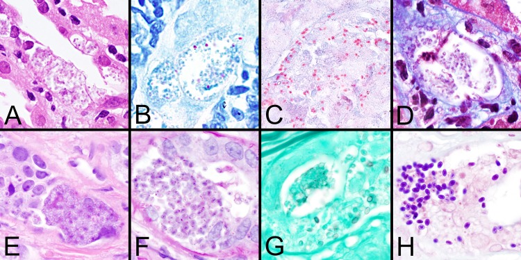
FIG 3.

Histologic staining patterns for microsporidial spores in tissue. (A to D) Images demonstrate spores of Encephalitozoon cuniculi in sections of kidney stained with H&E, Z-N, modified trichrome (Ryan blue), and Weber's modified trichome stain, respectively. Note that the spores are only focally positive with the Z-N acid-fast stain. (E to H) Images demonstrate the larger spores of Anncaliia algerae in eccrine skin glands stained with H&E, PAS, GMS, and Brown and Brenn (tissue Gram stain) stain, respectively. Note the dot-like PAS positivity and the focal staining with GMS. All images are shown at ×1,000 magnification.
