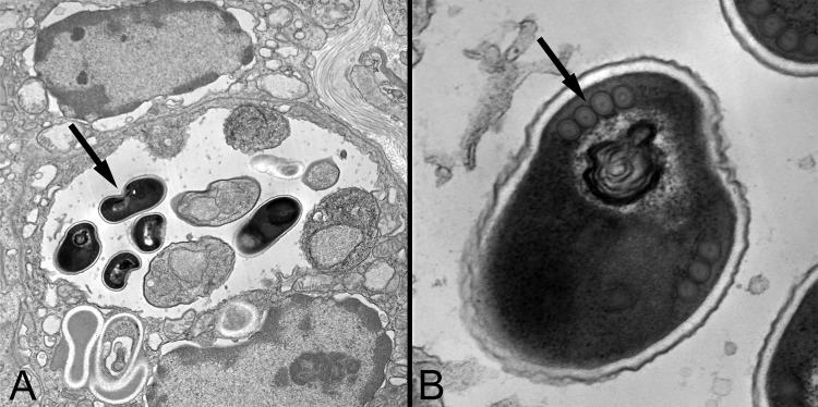
FIG 5.

Transmission electron micrographs of Encephalitozoon cuniculi organisms within a renal tubular cell. (A) Note that the dark spores (arrow) are within a parasitophorous vacuole, as is characteristic for Encephalitozoon spp. (×11,000 magnification). (B) On higher magnification (×68,000 magnification), a single row of polar filament cross sections is seen (arrow), which is also consistent with the diagnosis of Encephalitozoon spp. The final species determination was made by PCR.
