FIG 1.
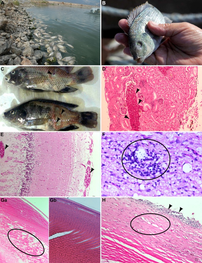
Characteristics of tilapia disease and pathological findings. Tilapia disease in commercial hybrid tilapia (O. niloticus × O. aureus hybrid) (A and C to E) and in wild tilapia (S. galilaeus) from the Sea of Galilee (B and F to H). (A) Tilapia disease outbreak in a commercial pond results in massive mortality (August 2013; courtesy of Nathan Wajsbrut). (B) Diseased tilapia demonstrating shrinkage of the eye and loss of ocular functioning (phthisis bulbi). (C) Gross pathology of skin includes multifocal to coalescing dermal erosions and ulcers (arrowheads; courtesy of Nathan Wajsbrut). (D) Kidney and interstitium. The arrowheads mark a dilated vein packed with large numbers of red blood cells (congestion). Hematoxylin and eosin (H&E) stain ×10. (E) Brain and cortex. The arrowheads mark dilated blood vessels packed with large numbers of red blood cells within the leptomeninges and gray and white matter. H&E stain ×10 was used. (F) Brain and cortex. Perivascular cuffs of lymphocytes (encircled). H&E stain ×40 was used. (Ga) Lens. Cataractous changes characterized by formation of eosinophilic spherical structures (morgagnian globules) accompanied by degeneration of crystalline fibers (encircled). H&E stain ×10 was used. (Gb) Control lens from healthy fish. H&E stain ×10 was used. (H) Eye and cornea. Loss of integrity of the overlying squamous epithelium with inflammatory infiltrate (arrowheads) and multiple capillaries within the stroma (neovascularization; encircled). The collagen fibers within the superficial stroma are smudged and are stained pale eosinophilic (corneal edema). H&E stain ×10 was used.

