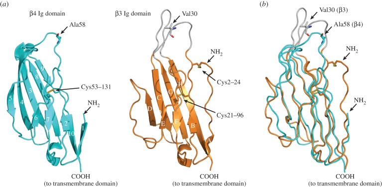
Figure 5.
The atomic resolution structure of the β4 subunit Ig domain. (a) The β4 Ig domain (PDB ID code 4MZ2) and comparison with similar orientation with the β3 Ig domain. (b) Supposition of the β4 Ig domain (cyan) and a single β3 Ig domain protomer (orange), showing the location of the Ala58 residue (corresponding to the free Cys58 residue of β4) and the Val30 residue of β3 mentioned in the text.

