
Figure 1. Autophagic puncta in hearts of mCherry-LC3 mice on chow or high fat diet (HFD).
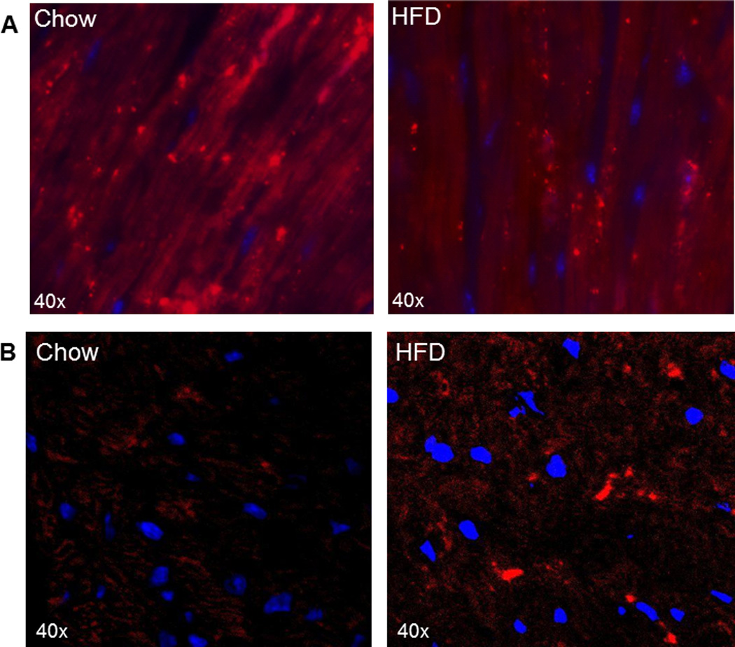
A. Representative heart sections of mice from the Mentzer study after 15 wk of chow or Teklad D12492 high fat diet. Cardiac sections (5–6 µm) were made using a cryostat, mounted on glass slides and nuclei stained with Hoechst 33342. Images were acquired with a Nikon TE300 fluorescence microscope equipped with a 60× Plan Apo objective with excitation/emission wavelengths of 560/630 nm. (Unpublished images generously provided by Dr. Bruce Ito and Dr. Robert Mentzer.) B. Representative heart sections of mice from the Abel study after 12 wk of chow or western diet. Images were taken with Zeiss LSM 510 confocal microscope using x63 immersion oil objective with excitation/emission wavelengths of 543/613 nm. (Unpublished images generously provided by Dr. Bharat Jaishy and Dr. E. Dale Abel.)