
Figure 2.
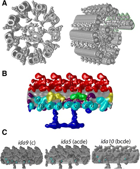
3D structure of Chlamydomonas flagella reconstructed by cryo-electron tomography and subtomogram averaging. (A) Structure of the entire axoneme by fitting averaged 96-nm periodic units to a tomogram. One MTD, discussed in the following sections, is enclosed by green lines in the right panel. (B) Averaged 96-nm unit. Red: outer dyneins. Cyan: inner dyneins. Blue: Radial spokes. Yellow: IC/LC of dynein f. Green: DRC. Grey: microtubule doublets (MTDs). Purple: unidentified density. Dynein isoforms were assigned based on (C). (C) Flagella structure of Chlamydomonas mutants used for identification of dynein isoforms. Missing IDA species are indicated.