Figure 4.

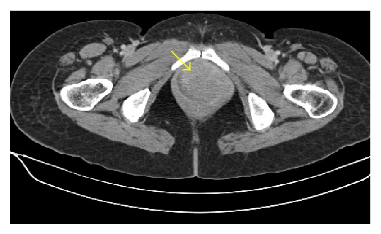
Computed tomography (CT) image of the pelvis shows well-defined homogenously enhancing lesion which is arising from left sided anterolateral wall of the vagina and projecting into vaginal lumen.

Computed tomography (CT) image of the pelvis shows well-defined homogenously enhancing lesion which is arising from left sided anterolateral wall of the vagina and projecting into vaginal lumen.