
Figure 1.
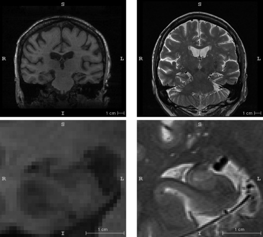
Example slices from the T1‐weighted (left) and T2‐weighted (right) images of the hippocampal region from one of the subjects in this study. The bottom panel is a zoomed in region around the right hippocampus. The T1‐weighed image is representative of what we describe as “routine” MRI in the text while the T2‐weighted image is an example of a “dedicated” MRI scan tailored for hippocampal subfield imaging. The slice plane is coronal for the T1‐weighted image and oblique coronal (orthogonal to the hippocampal main axis) for the T2‐weighted image.