
Figure 3.
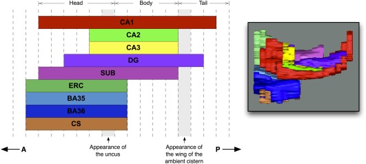
The extent along the anterior‐posterior axis (A–P in the figure) of the different anatomical labels included in the manual segmentation protocol. Dashed vertical lines outline MRI slices (the number of slices is variable from subject to subject). A 3D rendering of the manual segmentation viewed from a location superior to the hippocampus is shown for reference. Abbreviations: CA: cornu ammonis; DG: dentate gyrus; SUB: subiculum; ERC: entorhinal cortex; BA35/36: Brodmann area 35/36 (which together form the perirhinal cortex); CS: collateral sulcus. [Color figure can be viewed in the online issue, which is available at http://wileyonlinelibrary.com.]