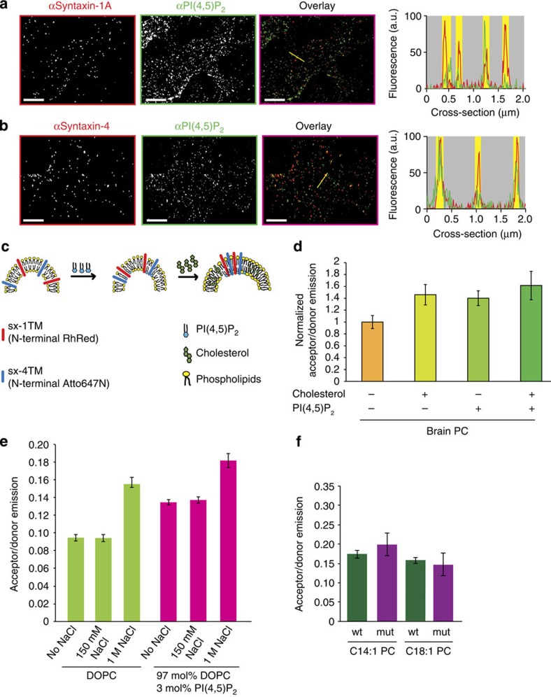
Figure 2. Ionic interactions and hydrophobic mismatch act synergistically on syntaxin clustering.
(a,b) Both syntaxin 1 and syntaxin 4 clusters colocalize with PI(4,5)P2. Plasma membrane sheets derived from PC12 cells were immunostained for PI(4,5)P2 (green), syntaxin 1 (a; red) and syntaxin 4 (b; red) and imaged by two-colour STED microscopy. The graphs show the fluorescence intensity profiles as indicated in the figures (syntaxin 1 and 4: red profiles; PI(4,5)P2: green profiles); yellow bars depict the positions of the domains. (c,d) Both PI(4,5)P2 and cholesterol enhance co-clustering of sx-1TM and sx-4TM. FRET was measured after reconstitution in large unilamellar vesicles containing sx-1TM labelled with Rhodamine Red (donor fluorophore) and sx-4TM with Atto647N (acceptor) composed of porcine brain PC without or with 1 mol% PI(4,5)P2 and/or 30 mol% cholesterol (± range from two independent reconstitutions, three technical repeats each). (e) Clustering of sx-1TM by FRET in the presence or absence of 150 mM or 1 M NaCl and in DOPC liposomes in the absence or presence of 3 mol% PI(4,5)P2. (f) Clustering of sx-1TM (green) and the sx-1TM oligomerization mutant (purple), monitored by FRET, in C14:1 PC or C18:1 PC liposomes. Error bars: range from two independent reconstitutions, three technical repeats each. Scale bars, 2 μm.

