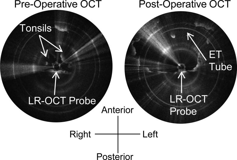
Figure 6.
Left: Pre-operative axial OCT image of the oropharynx of the 20.1kg 7-year-old male seen in Figure 5, showing the tonsils, which obscure the ET tube. Right: Post-operative axial OCT image of the oropharynx of the same patient. The tonsils have been removed, and the airway lumen is larger. Note the irregular appearance of the tissue border. Very bright white dots along the tissue surface indicate a thin film of blood and mucus.

