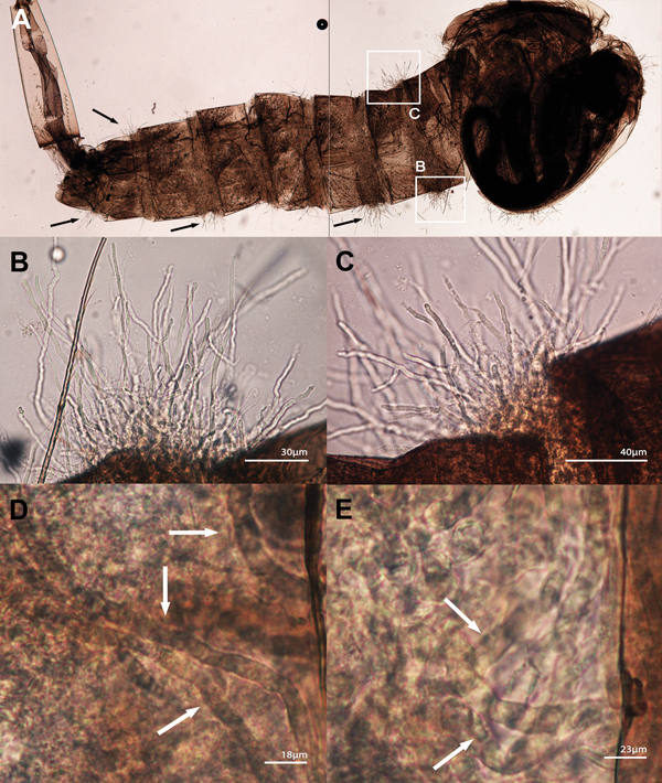
Figure 4.

Lagenidium giganteum from mammal experimental infection using Culex pipiens mosquito larvae. A) Composite of 2 photographs showing an instar 3 C. pipiens larvae infected with 1 of the 5 tested strains of L. giganteum recovered from dogs with lagenidiosis (MTLA01, type strain). Note the mycelioid structures emerging from the infected larvae (arrows). B, C) Enlargements of the 2 white boxes in (A) showing details of the mycelioid structures emerging between the segments of the larvae. D, E) Aggressiveness of the invading mycelioid structures (arrows) within the body of C. pipiens larvae.
