
Fig. 1. Impact of HDL dose on Aβ deposition and neurotoxicity.
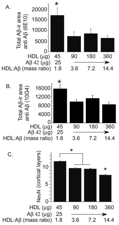
Aβ42 was icv infused at 25 μg/200 μl (0.2% DMSO) with different mass ratios of HDL. Rats were sacrificed at 4 weeks post-infusion and evaluated for Aβ deposits using monoclonal antibodies 6E10 (A) or 10G4 (B). Neuron nuclei staining in layer 2 of the entorhinal cortex was evaluated using NeuN (C). Values are shown as the mean ± SD. * p < 0.05 represent a significant difference between vehicle (HDL) and treatment (HDL + Aβ).