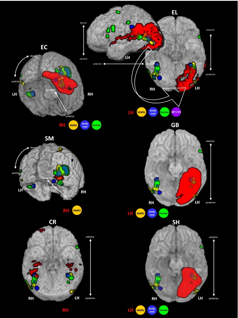
Fig. 3.
Situating the patients’ lesions with respect to visual regions standardly associated with biological motion processing, presented in rendered fashion. Lesion of each patient is delineated in red based on the structural images (Methods and Supporting Information). Regions consistently associated with biological motion processing are based on statistical maps of a meta-analysis (13): regions in blue are significantly activated to biological motion, regions in yellow are sensitive to static bodies, and regions in green are sensitive to human movement over nonhuman movements. As summarized in Table 3, ventral visual regions associated with all of the three types of biological motion perception were severely affected by brain damage in one or more of the ventral visual patients, including the ventral aspect of EBA (v-EBA, in EC, SM, and GB). This finding indicates that the spared perceptual thresholds for biological motion perception do not rely on the integrity of the ventral visual regions associated with biological motion processing. MT+/V5, middle temporal motion sensitive region; RH/LH, right/left hemisphere. See also Table 3.

