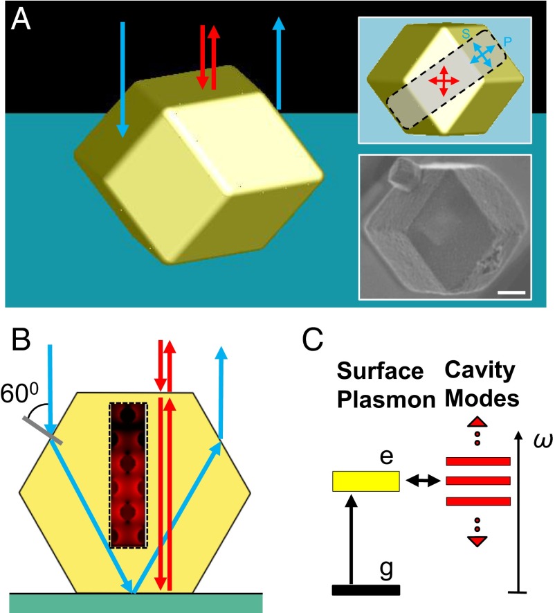
Fig. 1.
A polaritonic photonic crystal made by DNA-programmable assembly. (A) Three-dimensional illustration of a plasmonic PPC, in the shape of a rhombic dodecahedron, assembled from DNA-modified gold nanoparticles. Red arrows indicate light rays normal to the underlying substrate, impinging on and backscattering through a top facet of the crystal (FPMs). The blue ones represent light rays entering through the slanted side facets and leaving the PPC through the opposite side, not contributing to the FPMs (Fig. S2). The top right inset shows the top view of the crystal with two sets of arrows defining two polarization bases at the top and side facets. The bottom right inset shows an SEM image of a representative single crystal corresponding to the orientation of the top right inset. (Scale bar, 1 µm.) (B) A 2D scheme showing the geometric optics approximation of backscattering consistent with the explanation in A. The hexagon outline is a vertical cross-section through the gray area in the top right inset of A parallel to its long edge. The box enclosed by a dashed line depicts the interaction between localized surface plasmons and photonic modes (red arrows; FPMs) with a typical near-field profile around gold nanoparticles. The contribution of backscattering through the side facets (blue arrows) to FPMs is negligible. (C) Scheme of plasmon polariton formation. The localized surface plasmons (yellow bar) strongly couple to the photonic modes (red bars; FPMs).

