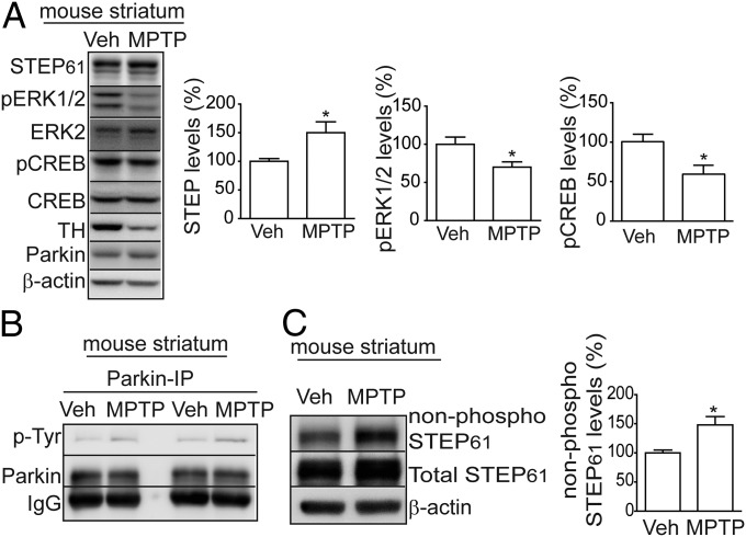
Fig. 5.
Striatal STEP61 levels are elevated after MPTP lesion. (A) Immunoblot and quantification indicate that STEP61 expression was increased and pERK1/2 phosphorylation and pCREB levels decreased in MPTP-treated mouse striatum compared with vehicle-treated controls. STEP61 levels were normalized to β-actin for quantification (n = 8; mean ± SEM; *P < 0.05; Student’s t test). (B) Representative immunoblot indicates that parkin tyrosine phosphorylation was increased in MPTP-treated striatal lysates, after immunoprecipitation of parkin and detection with a pan phospho-tyrosine antibody (n = 2). (C) Active, non-phospho-STEP61 and total STEP61 levels were increased in MPTP-lesioned mouse striatum compared with vehicle-treated controls. STEP61 levels were normalized to β-actin (n = 8; *P < 0.05; Student’s t test).

