Fig. 4.
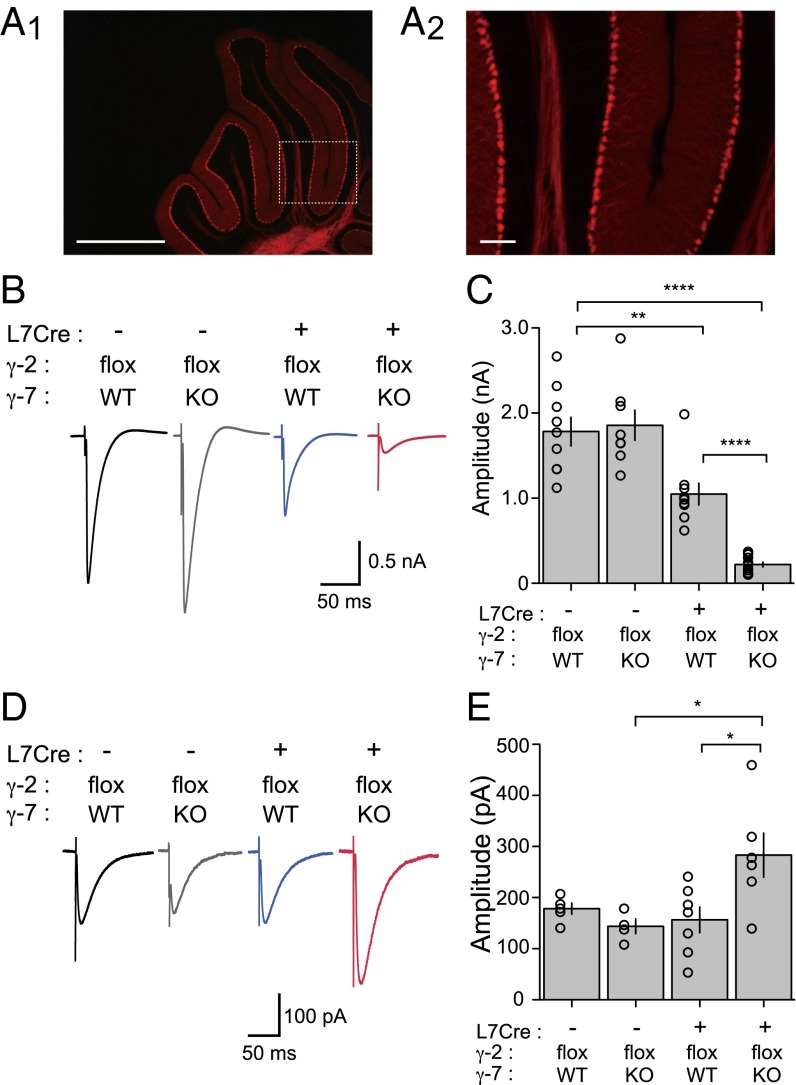
AMPA receptor-mediated synaptic currents were significantly reduced in Purkinje cell-specific γ-2/γ-7-double KO mice. (A) Fluorescent micrograph of cerebellum prepared from L7Cre; ROSA26-tdTomato mice. L7Cre mouse was crossed with ROSA26-tdTomato reporter line and analyzed at 2 mo old. White box in Left indicates brain region shown in Right. (Scale bars: Left, 1 mm; Right, 100 µm.) (B) Representative traces of cf-EPSCs recorded from γ-2 PC WT; γ-7 WT (L7Cre−; γ-2flox; γ-7 WT, black), γ-2 PC WT; γ-7 KO (L7Cre−; γ-2flox; γ-7 KO, gray), γ-2 PC KO; γ-7 WT (L7Cre+; γ-2flox; γ-7 WT, blue), and γ-2 PC KO; γ-7 KO (L7Cre+; γ-2flox; γ-7 KO, red) mice. (C) Summary bar graph showing peak amplitudes of cf-EPSCs recorded from P63 to 4-mo-old mice at the holding potential of −20 mV. (D) Representative traces of KAR-mediated currents recorded from γ-2 PC WT; γ-7 WT (black), γ-2 PC WT; γ-7 KO (gray), γ-2 PC KO; γ-7 WT (blue), and γ-2 PC KO; γ-7 KO (red) mice in the presence of 100 µM GYKI 53655 at −70 mV. (E) Summary bar graph showing the KAR EPSCs. Open circles represent values from individual cells. Asterisks indicate significance, *P ≤ 0.05, **P ≤ 0.01, ****P ≤ 0.0001.

