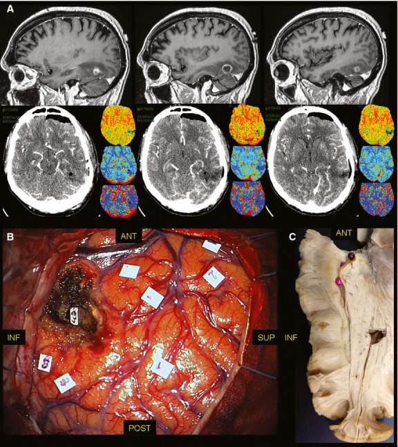
Fig. 2.

(A) On the upper line, the pre-operative sagittal T1 contrast-enhancing MRI shows a HGG with a nodular enhancing lesion surrounded by hypointense tissue. On the lower line, the post-operative perfusion CT-scan demonstrated the resection of the lesion. (B) The DES allowed to push the resection beyond the non-functional nodular enhancing tissue up to the identification of the inferior limit of the OR (tag 43), at the inferior margin of the lateral wall of the trigone, such as demonstrated in the anatomical cadaver dissection (C) with Klingler's technique. CT, computer tomography; DES, direct electrical stimulation; HGG, high-grade glioma; MRI, magnetic resonance imaging; OR, optic radiation.
