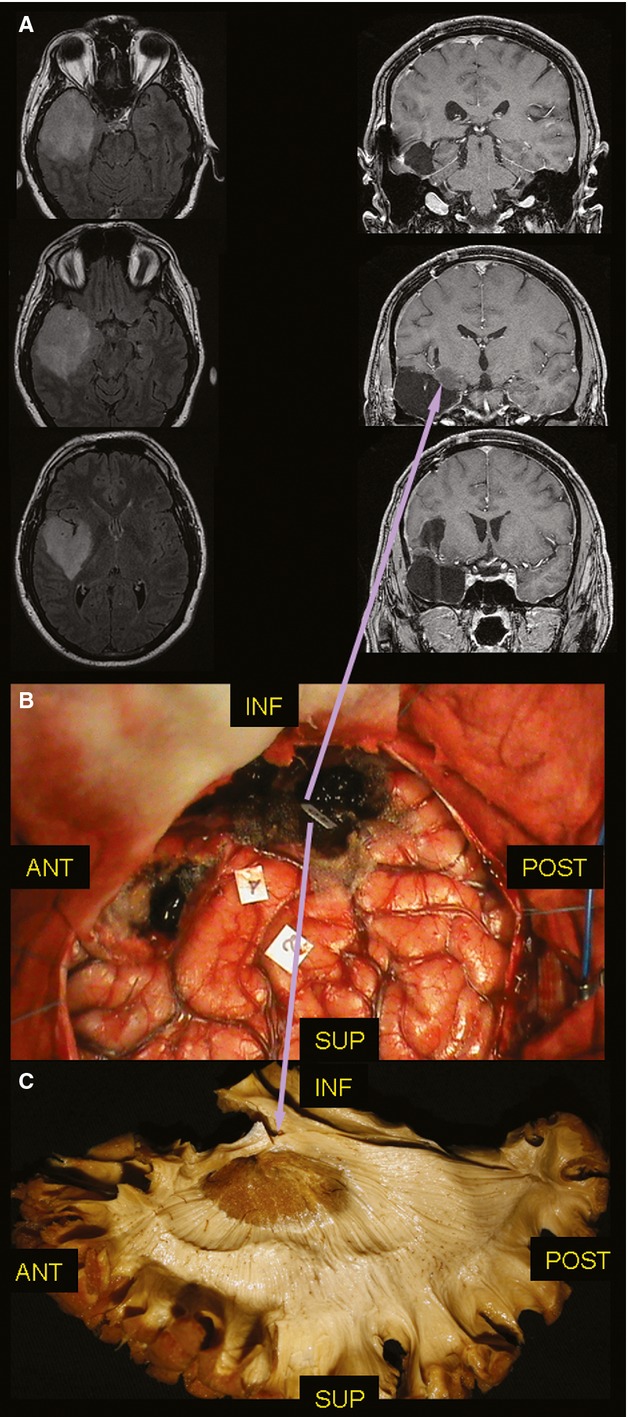
Fig. 3.

(A) On the left side, the pre-operative axial FLAIR MRI shows a diffuse lesion infiltrating the whole temporal lobe and the insula. On the right side, the post-operative coronal T1 MRI confirmed the limits of resection, at the level of the TS. (B) The DES allowed the resection of the temporal lobe and of the entire insula with a 6 cc residual tumour within the temporal stem. (C) The stimulation site of the superior OR (tag 40) matches (inferior purple arrow) with the course of these fibres at the level of the temporal stem identified with post-mortem dissection of the WM and the residual tissue at the early post-operative MRI (superior purple arrow). DES, direct electrical stimulation; MRI, magnetic resonance imaging; OR, optic radiation; TS, temporal stem; WM, white matter.
