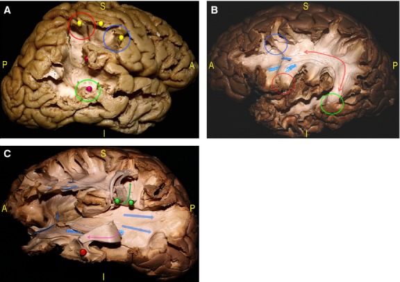
Fig. 5.

(A) The dissection started with the resection of the posterior MTG demonstrating the posterior indirect component of the SLF, connecting the Wernicke's territories (red pin, green circle) to the Geschwind territories within the inferior parietal lobule (IPL; red pin), and the anterior indirect component, connecting the Geshwind territories to the posterior IFG (yellow, pins, red and blue circles). (B) Deeper the AF (red arrow) is shown connecting the IFG (red circle) and MFG (blue circle) to the STG, MTG and ITG (green circle). (C) The AF and its termination are then cut and turned up (green arrow, green pins), and the ILF is detached and turned anteriorly (red pin, pink arrow) to show the deeper layer of the SS composed of the IFOF and superior and inferior components of the OR (blue arrows). AF, arcuate fasciculus; IFG, inferior frontal gyrus; IFOF, inferior fronto-occipital fascicle; ILF, inferior longitudinal fascicle; IPL, inferior parietal lobule; ITG, inferior temporal gyrus; MFG, middle frontal gyrus; MTG, middle temporal gyrus; OR, optic radiation; SLF, superior longitudinal fascicle; SS, stratum sagittalis.
