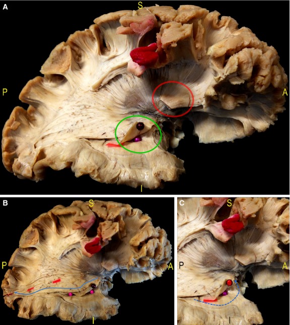
Fig. 7.

(A) After cutting the IFOF stem at the level of the ventral EC (red circle) and removal of the fibres of the occipito-temporal portion of the IFOF, the deeper putamen and the fibres of the anterior OR are shown (green circle). (B) In particular, the fibres of the inferior and superior component were separated by means of red tags, starting posteriorly from the calcarine sulcus. The inferior OR was followed on its anterior course along the temporal horn (blue arrow, pink pins) with the demonstration of the fibres Meyer's Loop, enveloping in an antero-medial direction the tip of the temporal horn. (C) The dissection allowed to lift up the main component of the Meyer's Loop without cutting in order better demonstrate its relationships with the tip of the temporal horn (red pin, blue arrow). EC, external capsule; IFOF, inferior-fronto-occipital fascicle; OR, optic radiation.
