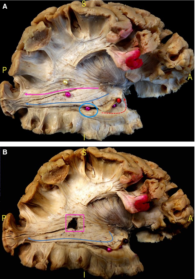
Fig. 8.

(A) The border between inferior and superior OR is highlighted starting posteriorly from the calcarine sulcus and it corresponds to the inferior margin of the lateral wall of the trigone (pink pin). The superior OR completely covers the lateral wall of the trigone and then courses antero-inferiorly within the posterior thalamic radiation (pink arrow). The inferior OR has a more inferior course along the lateral and inferior wall of the ventricle (blue circle, inferior intra-ventricular pink pins) up to the enlarge in an antero-medial direction (blue arrow) around the tip of the temporal horn (Meyer's Loop, lifted up by a red pin). (B) After cutting the fibres between the two pins in picture (A), the trigone is opened (pink square), demonstrating the course of the inferior OR at its inferior margin (blue arrow). OR, optic radiation.
