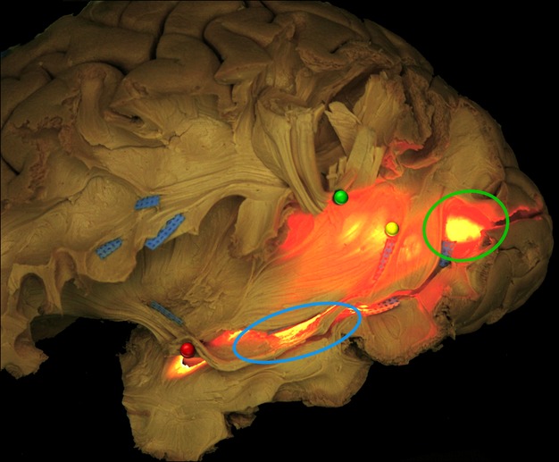
Fig. 9.

In this figure, a transventricular illumination is applied from the mesial side of a left hemisphere. It allowed to better show the relationships of WM fibres isolated during the dissection with the ventricular structures. The more superficial WM fibres, particularly AF and ILF, are, respectively, cut and lifted up (green pin) and lifted ‘in situ’ highlighted with blue tag. The more posterior portions of IFOF and OR before the occipital terminations are shown overlapping the occipital horn (green circle) and running anteriorly. The inferior and superior components of OR and IFOF course all along the lateral ventricle (yellow pin), crossing the ILF main stem (blue tags), up to the tip of the temporal horn (blue circle) where the fibres of the Meyer's Loop are lifted up with a red pin. AF, arcuate fasciculus; IFOF, inferior fronto-occipital fascicle; ILF, inferior longitudinal fascicle; OR, optic radiation; WM, white matter.
