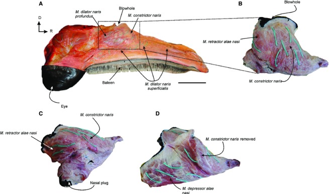
Fig. 7.

Nasal muscles of Eubalaena australis: (A) right lateral view of the head of a neonate specimen showing the most superficial nasal muscles in situ; (B) lateral and (C,D) caudo-lateral view of the nasal muscles removed from the nasal fossa showing the fibers directions (green threads) of the m. contrictor naris m. retractor alae nasi and m. depressor alae nasi. D, dorsal; R: rostral. Scale bar: 15 cm (A).
