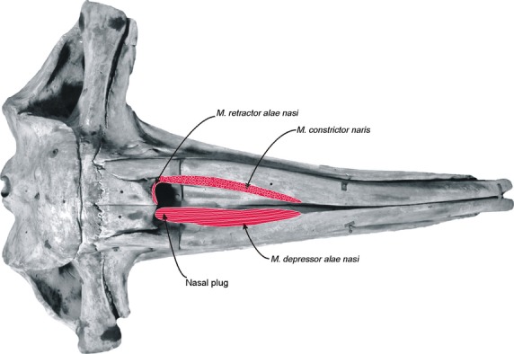
Fig. 9.

Schematic reconstruction of the nasal muscles of Eubalaena australis (skull in dorsal view). In the left side of the skull are represented the attachments of the most superficial nasal muscles, the m. constrictor naris and m. retractor alae nasi. Note that the fibers of the m. constrictor naris are sectioned in a horizontal plane with respect to the sagittal plane of the skull. In the right side of the skull is represented the attachment of the deepest nasal muscle, the m. depressor alae nasi, along the whole ventral surface of the nasal fossa.
