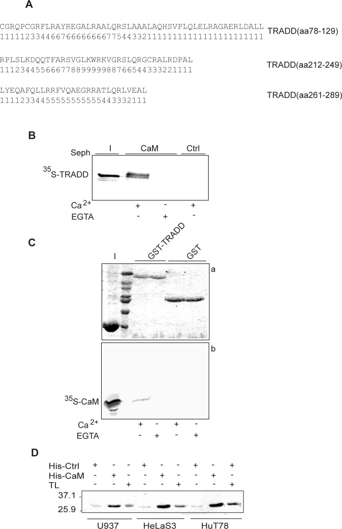
Figure 1. Ca2+-dependent binding of CaM to TRADD.
A: CaM target database analysis. Amino acid sequences of the predicted CaM binding sites in human TRADD are shown along with the corresponding probability scores. B: autoradiography of CaM pull-down assay. (I) shows input of 35S-TRADD (∼ 33 kDa) incubated with CaM sepharose (CaM) or control sepharose (Ctrl) beads in Ca2+ or EGTA binding buffer. C: autoradiography of pull-down assay of 35S-CaM with GST or GST-TRADD. Panel a) shows a 12% SDS-PAGE stained with coomassie and panel b) the corresponding autoradiogram for 35S-CaM. D: western blot of His-CaM pull-down assays. His-CaM or His-Ctrl (control, cyclophilin) bound to Ni-NTA agarose beads were incubated with cell lysates, as indicated. TL indicates total cell lysates. Positions of the molecular weight standards are indicated. The data shown are representative of at least three independent experiments.

