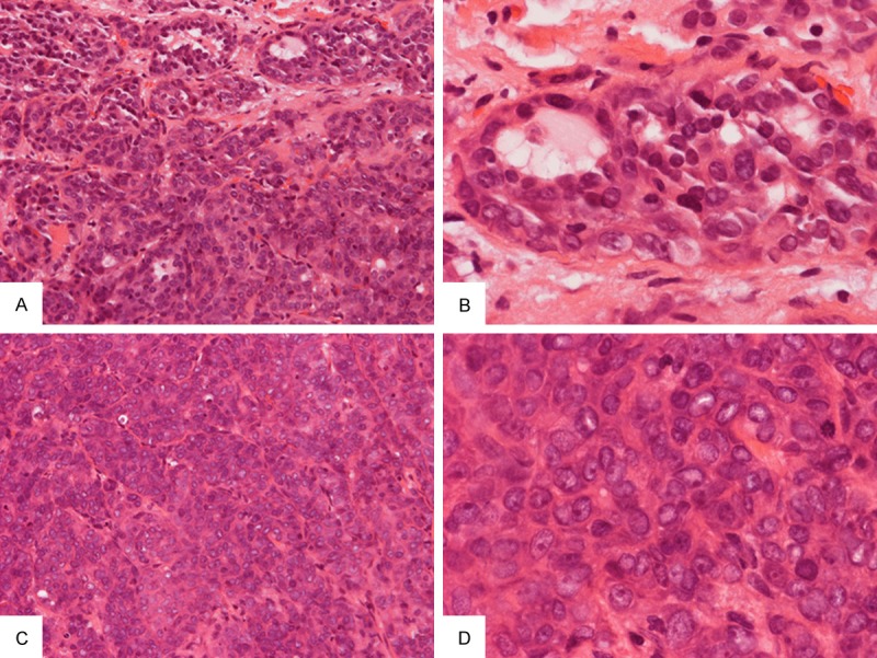
Figure 2.

Biopsy specimen: A few small neoplastic glands with pale eosinophilic secretion are present at the periphery of the tumor (A) and the tumor cells have small to medium sized nuclei without prominent nuclei (B). The bulk of the neoplastic component is composed of solid cords and trabeculae of cells separated by thin fibrous septa (C). The nuclei are enlarged and contain prominent nucleoli (D) in contrast to those with gland formation as illustrated in (B). Original magnification in (A) and (C) is 20×, in (B) and (D) is 60×.
