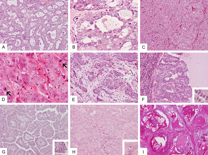
Figure 3.

Resection specimen: Areas with neoplastic gland formation (A) are noted mainly at the periphery of the tumor. These glands contain small amount of pale eosinophilic secretion (B). The bulk of the tumor is solid and is composed of cords or nests of cells with high-grade nuclei with prominent nucleoli separated by thin fibrous septa with delicate blood vessels (C and D). Multiple mitoses are present (arrows in D). Infiltrating tumor with desmomplastic reaction is also noted (E). Histology of the metastasis to cervical lymph nodes includes cystic (F) growth pattern (G) with low-grade nuclei (inset in F), papillary growth pattern (G) with low-grade nuclei (inset in G), and solid areas (H) with high-grade nuclei (inset in H). Secretions in the areas are strongly positive for PAS stain (I). Original magnification in (A, C, E-I) and is 20×; in (B), (D), and insets in (F), (G), and (H) is 60×.
