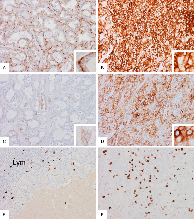
Figure 4.

Immunohistochemistry: Expression of cytokeratin 7 is patchy in areas with gland formation (A) and is mostly found at the base of the lining cells (inset in A) but the expression is diffuse in solid areas (B). Expression of cytokeratin 8/18 is only focal in areas with gland formation (C) but is wide spread in solid areas (D). In the metastasis, the Ki67 labeling index is low in the papillary area (E) but high in the solid areas (F). Original magnification in all panels is 20× and in all insets is 60×.
