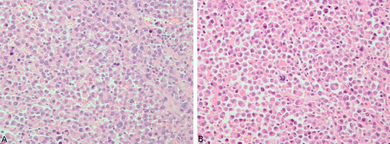
Figure 2.

A. Microscopy showed that the mass was composed of atypical plasmacytoma cells with wheel-spoke-like nucleus, clusters of plasma cells with eccentric nuclei (Hematoxylin-eosin staining; original magnification, 400×). B. Occaslonal bi- and multi-nucleation and 1-2 mitotic figures in the nuclei were observed in high-power field microscopy (Hematoxylin-eosin staining; original magnification, 400×).
