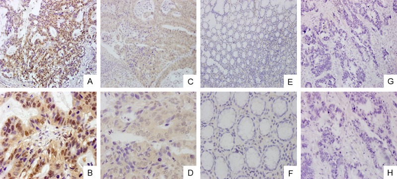
Figure 2.

Immunohistochemical analysis of ECT2. A, B. ECT2 was strongly expressed in gastric cancer tissues; magnifications were × 200 and × 600, respectively. C, D. ECT2 was weakly expressed in gastric cancer tissues; magnifications were × 200 and × 600, respectively. E, F. ECT2 negative in normal gastric tissues; magnifications were × 200 and × 600, respectively. G, H. negative sample for immunostaining of ECT2, with phosphate-buffered saline replacing primary antibody against ECT2; magnifications were × 200 and × 600.
