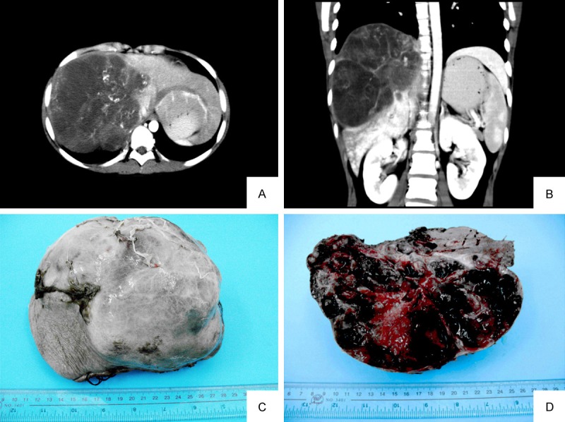
Figure 1.

A, B. CT scan showed a hypodense mass with a multicystic appearance in the right lobe liver. Peripheral rim of the mass was enhanced but not the hypodense area in enhanced scan (Case No. 8). C. The tumor was globular and well demarcated with complete encapsulation (Case No. 9). D. The cut-surface is grey-white, with prominent haemorrhagic and necrotic areas.
