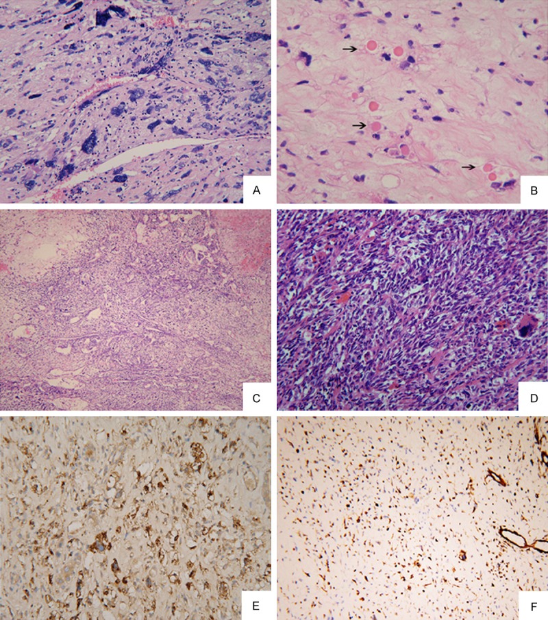
Figure 2.

(A) There were foci of uniform small round cells admixed with a small number of pleomorphic cells in an edematous background (HE × 200) (Case No. 4); (B) Variable sized eosinophilic globules were present in the cytoplasm of large tumor cells or in extracellular matrix (arrow) (HE × 200); (C) Haemangiocytoma-like compact areas with collagenization (HE × 100) (Case No. 2); (D) Fibroblast-like fascicles and bundles were seen in compact areas (HE × 200) (Case No. 8); (E, F) Tumor cells were strongly positive for AAT (E) and Vimentin (F) (Case No. 4) (Envision Immunohistochemistry, × 100).
