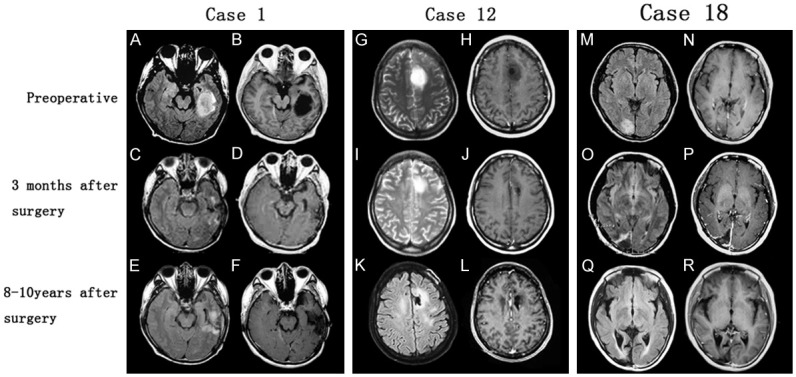
Figure 1.

Radiological findings in three cases of IDLGGs. Preoperative MRs incidentally revealed left temporal lesion (Case 1), left frontal lesion (Case 2) and right occipital lesion (Case 18). Postoperative MRs 3 months after operations demonstrated surgical changes with no obvious tumor residual and latest follow-up MRs showed no signs of tumor recurrence. (A, C, E, M, O, Q: axial FLAIR sequences; G, I, K: axial T2-weighted sequences; B, D, F, H, J, L, N, P, R: axial T1-weighted sequences after contrast enhancement). FLAIR = fluid attenuation inversion recovery.
