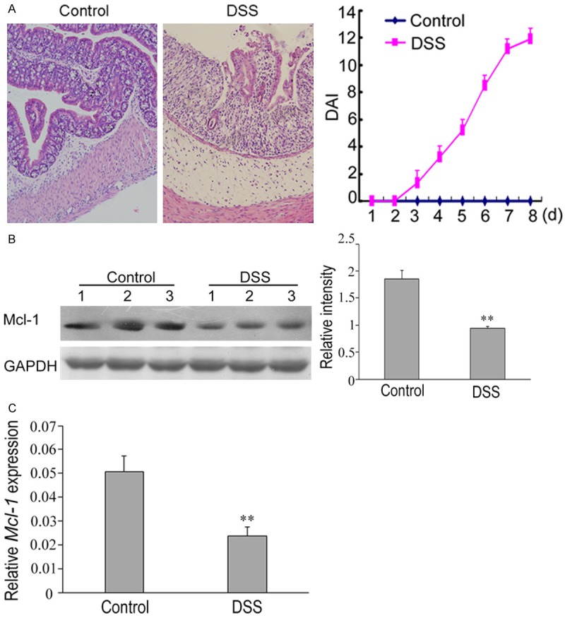
Figure 2.

Down-regulation of Mcl-1 in mice with DSS-induced experimental colitis. A. Histopathological changes in colon tissues of mice with DSS-induced colitis. Compared to the normal colon tissues, the epithelial structure of the colon tissues of mice with DSS-induced colitis was destroyed and a large number of lymphocytic infiltration was observed in the mucosa and submucosa. The disease activity index (DAI) was significantly higher in mice with DSS-induced colitis than in the control animals (right). B. The expression levels of mcl-1 in each group. The expression of mcl-1 protein was significantly decreased in mice with DSS-induced colitis compared to the control animals. Western blot analyses of band intensity from three independent experiments are presented as the relative ratio of mcl-1 to β-actin. **P < 0.01 vs. control. C. RT-qPCR analysis of the relative expression of miR-29a in the control and DSS group. U6 served as an internal control. **P < 0.01 vs. control.
