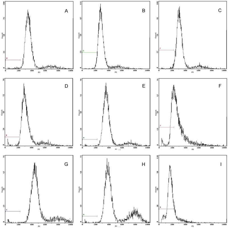
Figure 2.

Flow cytometry to detect cell apoptosis in three groups. A-C. Control group at 2 weeks, 4 weeks and 8 weeks. The rate of apoptosis cell was 1.85%, 1.91% and 1.79% respectively. D-F. Suture group at 2 weeks, 4 weeks and 8 weeks after surgry. The rate of apoptosis cell was 2.07%, 2.32% and 34.8% respectively. G-I. Model group at 2 weeks, 4 weeks and 8 weeks after surgry. The rate of apoptosis cell was 2.26%, 2.47% and 81.10% respectively.
