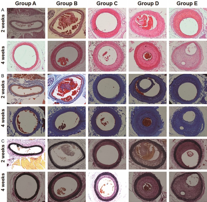
Figure 6.

Histopathological investigation of CCA segments on days 14 and 28. Representative microscopic images of tissue sections from CCA segments in rat models on days 14 and 28 after treatment. Blood clots and their organization in these tissue sections were identified using hematoxylin & eosin, Masson trichrome, and Elastica van Gieson (magnification: 100×).
